MEF2D
MEF2D,即肌细胞增强因子2D,别名Myocyte enhancer factor 2D。它是MADS盒家族的成员之一,参与多种细胞生命活动的调控。它通过与DNA上的MEF2结合位点相互作用,调控下游基因的转录,影响细胞增殖、分化、迁移和凋亡等过程。MEF2D在肌肉生成、神经系统发育、肝纤维化以及肿瘤发展中扮演重要角色。特别是在肿瘤生物学中,MEF2D的异常表达与多种肿瘤的侵袭性、转移能力和患者预后密切相关,使其成为癌症治疗的潜在靶点。
热销产品
MEF2D Antibody (CSB-PA613522LA01HU)
验证数据
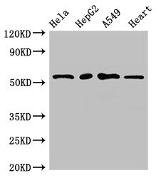
Western Blot
Positive WB detected in: Hela whole cell lysate, HepG2 whole cell lysate, A549 whole cell lysate, Mouse heart tissue
All lanes: MEF2D antibody at 2.7µg/ml
Secondary
Goat polyclonal to rabbit IgG at 1/50000 dilution
Predicted band size: 56, 57, 51, 50 kDa
Observed band size: 56 kDa
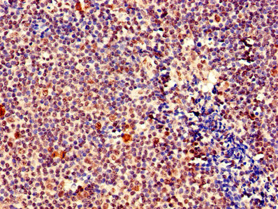
IHC image of CSB-PA613522LA01HU diluted at 1:400 and staining in paraffin-embedded human tonsil tissue performed on a Leica BondTM system. After dewaxing and hydration, antigen retrieval was mediated by high pressure in a citrate buffer (pH 6.0). Section was blocked with 10% normal goat serum 30min at RT. Then primary antibody (1% BSA) was incubated at 4°C overnight. The primary is detected by a biotinylated secondary antibody and visualized using an HRP conjugated SP system.
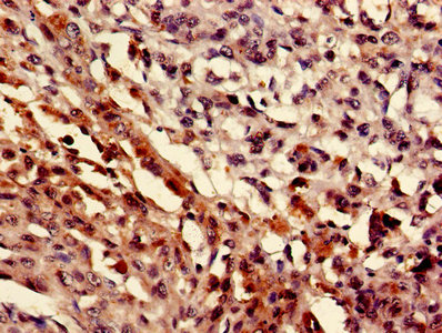
IHC image of CSB-PA613522LA01HU diluted at 1:400 and staining in paraffin-embedded human melanoma performed on a Leica BondTM system. After dewaxing and hydration, antigen retrieval was mediated by high pressure in a citrate buffer (pH 6.0). Section was blocked with 10% normal goat serum 30min at RT. Then primary antibody (1% BSA) was incubated at 4°C overnight. The primary is detected by a biotinylated secondary antibody and visualized using an HRP conjugated SP system.
MEF2D Antibodies
MEF2D for Homo sapiens (Human)
| 产品货号 | 产品名称 | 种属反应性 | 应用类型 |
|---|---|---|---|
| CSB-PA010173 | Phospho-MEF2D (S444) Antibody | Human,Mouse,Rat | WB, IHC, ELISA |
| CSB-PA010174 | MEF2D Antibody | Human,Mouse,Rat | IHC, ELISA |
| CSB-PA613522LA01HU | MEF2D Antibody | Human, Mouse | ELISA, WB, IHC |
| CSB-PA613522LB01HU | MEF2D Antibody, HRP conjugated | Human | ELISA |
| CSB-PA613522LC01HU | MEF2D Antibody, FITC conjugated | Human | |
| CSB-PA613522LD01HU | MEF2D Antibody, Biotin conjugated | Human | ELISA |
MEF2D Proteins
MEF2D Proteins for Rattus norvegicus (Rat)
| 产品货号 | 产品名称 | 来源 |
|---|---|---|
| CSB-YP013674RA CSB-EP013674RA CSB-BP013674RA CSB-MP013674RA CSB-EP013674RA-B |
Recombinant Rat Myocyte-specific enhancer factor 2D (Mef2d) | Yeast E.coli Baculovirus Mammalian cell In Vivo Biotinylation in E.coli |
MEF2D Proteins for Homo sapiens (Human)
| 产品货号 | 产品名称 | 来源 |
|---|---|---|
| CSB-YP613522HU CSB-EP613522HU CSB-BP613522HU CSB-MP613522HU CSB-EP613522HU-B |
Recombinant Human Myocyte-specific enhancer factor 2D (MEF2D) | Yeast E.coli Baculovirus Mammalian cell In Vivo Biotinylation in E.coli |
MEF2D Proteins for Mus musculus (Mouse)
| 产品货号 | 产品名称 | 来源 |
|---|---|---|
| CSB-YP733979MO CSB-EP733979MO CSB-BP733979MO CSB-MP733979MO CSB-EP733979MO-B |
Recombinant Mouse Myocyte-specific enhancer factor 2D (Mef2d) | Yeast E.coli Baculovirus Mammalian cell In Vivo Biotinylation in E.coli |